
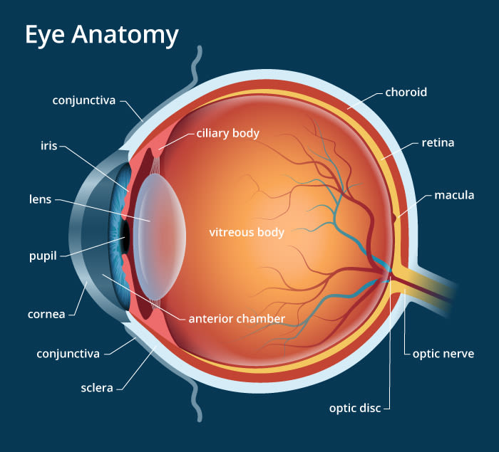
The first thing it touches is a thin veil of tears on the surface of the eye. Behind this is your eye’s front window, the cornea. This clear layer helps focus the light.
On the other side is liquid called the aqueous humor. It circulates throughout the front part of your eye and keeps pressure inside constant
After the aqueous humor, light passes through the pupil. This is the central round opening in your iris, the colored part of your eye. It changes size to control how much light gets in further back. 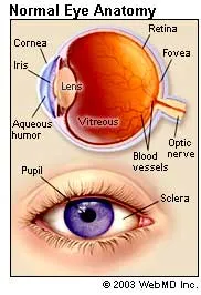 Next up is the lens. It works just like a camera to focus light. It adjusts shape depending on whether the light reflects off something near to you or far away.
This light now pierces the center of the eyeball. It’s bathed in moisture from a clear jelly known as the vitreous.
Unlike a movie screen, the retina has many parts:
Blood vessels bring nutrients to your nerve cells.
The macula is the bull’s-eye at the center of your retina. The dead center is called the fovea. Because it’s the focal point of your eye, it has more special, light-sensitive nerve endings, called photoreceptors, than any other part.
Photoreceptors come in two kinds: rods and cones. They’re special nerve endings that convert the light into electrochemical signals.
Retinal pigment epithelium (RPE) is a layer of dark tissue beneath the photoreceptors. These cells absorb excess light so the photoreceptors can give a clearer signal. They also move nutrients to (and waste from) the photoreceptors to the choroid.
The choroid is separate from the RPE. It lies behind the retina and is made up of many fine blood vessels that supply nutrition to the retina and the RPE.
Signals from the photoreceptors travel along nerve fibers to the optic nerve. It sends the signals to the visual center in the back of the brain.
And that’s how you see: Light, reflected from an object, enters the eye, gets focused, is converted into electrochemical signals, delivered to the brain, and is interpreted, or “seen,” as an image.
Source:- webmd